
Genetic engineering & the next pandemic
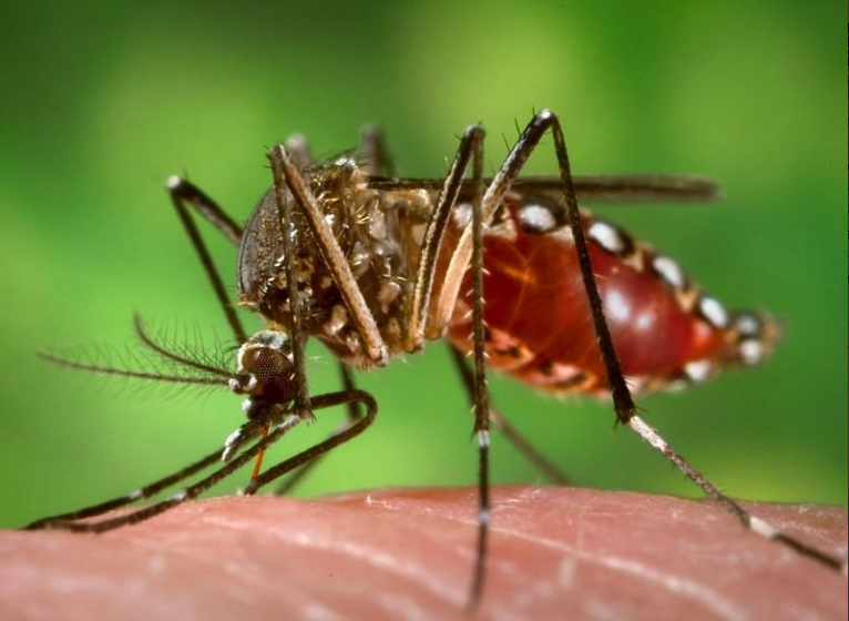
“For decades, the disease transmitted by the Aedes genus of mosquito slumbered, affecting mainly monkeys. In humans, Zika occasionally caused a mild disease of low concern…The situation today is dramatically different. Last year, the virus was detected in the Americas, where it is now spreading explosively. As of today, cases have been reported in 23 countries and territories in the region. The level of alarm is extremely high.” World Health Organization Director-General Dr. Margaret Chan
Once again, the mainstream media vultures are purposefully exploiting the latest (potentially global) health crisis threatening our communities, with the stampeding Zika Virus, ramping up their rhetoric and fear-mongering headlines.
The origins, and the nature of this outbreak, an unprecedented spike in cases of microcephaly associated with symptoms of the strain, occurring mainly in concentrated areas, seemingly without any environmental or biological catalyst, have left the entire medical community scratching their heads.
So what is actually behind this multi-faceted mystery? The answers are to be found on the ground, in the heart of these impacted communities. You can be certain your Government Health Department has a vested interest in withholding the truth from ever reaching the light of day.
Given the implications, and the financial fallout, not to mention the impact on countless families a cover-up of this magnitude is having, and will have, for generations, who can blame them? We’re now entering uncharted territory.
The Zika Virus (an RNA virus containing 10,794 nucleotides encoding 3,419 amino acids) has long been considered a relatively innocuous, albeit contagious, localized pathogen, typically producing Flu-like symptoms in the host, on average subsiding within one to two weeks; certainly nothing on the scale comparatively, to that of the Dengue or Yellow Fever Virus variety, let alone Ebola – each of which are associated with varying degrees of internal “blackish” bruising & widespread hemorrhaging (‘gastrointestinal bleeding, haematuria, skin petechiae, ecchymoses’) followed by rapid systemic deterioration, marked by Kidney failure, often leading to death.
‘Zika virus is an emerging mosquito-borne (Fla)virus that was first identified in Uganda in 1947 in rhesus monkeys through a monitoring network of sylvatic yellow fever. It was subsequently identified in humans in 1952 in Uganda and the United Republic of Tanzania. Outbreaks of Zika virus disease have been recorded in Africa, the Americas, Asia and the Pacific.‘
‘Most reports describe a self-limiting febrile illness that could easily be mistaken for another arboviral infection, such as dengue or chikungunya fever.` National Institutes of Health
Symptomatology commonly associated with Zika Virus includes the following:
•The incubation period ranges between approximately 3 to 12 days after the bite of an infected mosquito.
•Most of the infections remain asymptomatic (between 60 to 80%).
•Disease symptoms are usually mild and the disease is usually characterised by a short-lasting self-limiting febrile illness of 4–7 days duration without severe complications, with no associated fatalities and a low hospitalisation rate.
•The main symptoms are macular or papular rash, fever, arthralgia, non-purulent conjunctivitis/conjunctival hyperaemia, myalgia and headache. The maculo-papular rash often starts on the face and then spreads throughout the body. Less frequently, retro-orbital pain and gastro-intestinal signs are present.
However, until now, the epidemiology of Zika has demonstrated no consistent connection, whatsoever, to birth defects in humans. Clearly, some vital component must have changed in the composition (nucleotide sequencing) of the virus, to render it more virulent; a sudden, dramatic shift in its response/adaptability mechanism to the environment (or a marked deterioration in our capacity to respond/adapt to it), by which to explain this disturbing anomaly. Notwithstanding those options, a rogue element would have to have been introduced in order to disturb the balance.
The majority, if not all emerging viruses in the spectrum, have undergone so many changes, through decades of unmonitored laboratory experimentation, re-engineering, chemical synthesis and genetic replication via vaccine programs (ie. Polio, MMR), they no longer resemble anything close to the naturally occurring, “wild strains” previously found in the environment.
Science has proven that vaccines inhibit the immune response, particularly in babies and young children, bypass/breach their under-developed, naturally aligned defences (80% of which are situated in the Mucosal membrane and the gastro-intestinal tract/gut), attacking the newly formed primary core circuits – comprising the Blood-Brain barrier, Myelin sheath & Meninges. The methodology, justifying vaccine uptake, is fundamentally flawed.
“But from all of the other studies of toxic substances, the earlier you work with the central nervous system, the more likely you are to run into a sensitive period for one of these effects, so that moving from one month or one day of birth to six months of birth changes enormously the potential for toxicity. There are just a host of neurodevelopmental data that would suggest that we’ve got a serious problem. The earlier we go, the more serious the problem.” Dr. Bill Weil/Pediatrician, Academy of Pediatricians Committee on Environmental Health, CDC Simpsonwood Transcripts, 2000
How does this specific vulnerability to vaccines correlate with pregnant women?
Mother & child share the same immunity while the baby is ‘In Utero’ (all 3 trimesters); including the entire duration of breast-feeding after birth. The Placenta and breast milk (Colostrum) are inextricably linked, providing a baby’s primary initial source of nourishment through the long journey of formation in utero – while supplying the basic building blocks of life necessary to guarantee a safe transition into early childhood development.
Via this intrinsic lifeline, the fetus is also rendered highly susceptible to what are termed ‘endogenous retroviruses’ (remnants of ancestral exogenous retroviral infections fixed in the germ-line DNA), passed on inter-generationally – primarily the result of previous exposure (grand-parent to parent to offspring) to the Polio, Tdap & MMR vaccines, from the mid 1950’s through the late 1960’s. The infant inherits any repository of cross-contaminated residue found in these shots, left embedded in the organs, bound to genetic material.
It goes without saying that pregnant women are at a heightened risk of adverse reactions to vaccines.
‘To address this serious situation (Microcephaly linked to Zika Virus), in late 2014, the Ministry of Health announced the introduction of the Tdap vaccine (comprising Diphtheria, Tetanus & Pertussis toxoids) for all pregnant women in Brazil.‘
Granted, any widespread immunization campaign targeting pregnant women which correlates with the epicenter of a sudden surge (mainly throughout sectors of north-eastern Brazil) is highly suspicious, given the verifiable track-record of serious adverse (auto-immune) reactions, neurological trauma and neuro-developmental difficulties, occurring in babies and young children following vaccination with Tdap.
Surprisingly, in this instance, the timeline simply doesn’t match up. Here is why.
First of all, the Tdap vaccine was not strictly enforced as a preventative measure for pregnant women, the key word being “recommended”. Hence, the statistical numbers of those vaccinated women in this community cannot be fully verified.
More importantly, microcephaly has a unique signature which rules out any damage done beyond the second trimester.
‘Microcephaly is a rare neurological condition in which an infant’s head is significantly smaller than the heads of other children of the same age and sex. Sometimes detected at birth, microcephaly usually is the result of the brain developing abnormally in the womb or not growing as it should after birth.
Microcephaly can be caused by a variety of genetic and environmental factors. Children with microcephaly often have developmental issues. Generally there’s no treatment for microcephaly, but early intervention with supportive therapies, such as speech and occupational therapies, may help enhance your child’s development and improve quality of life.‘ Mayo Clinic
The brain and skull cavity develop throughout the first and second trimester. By 24 weeks gestation (in utero), the overall structure, mass & density of both areas has been firmly established.
‘The frontal bone, located superiorly and anteriorly on the skull, is the first of the cranial bones to begin ossification. The process begins to develop from two centers and is visible microscopically by alizarin-stain as early as 6-7 weeks gestation and radiographically by the 13th week.
There are two parietal bones located on each side of the brain, posteriorly to the frontal bone. The fetal parietals form from two centers of ossification that can be microscopically identified with alizarin-stain by 7-8 weeks gestation. Ossification of the two centers fuse quickly, forming an hour-glass shape in the early stages and rapidly develop into an ellipsoid recognizable by radiographic measures at around 20 weeks gestation. Although ossification and development begins at an early stage, the parietals are not identifiable by angles and borders until around the 24th week.’ Georgia State University, Department of Anthropology
‘The fifth week of pregnancy, or the third week after conception, marks the beginning of the embryonic period. This is when the baby’s brain, spinal cord, heart and other organs begin to form.
Seven weeks into your pregnancy, or five weeks after conception, your baby’s brain and face are rapidly developing.
By the 10th week of pregnancy, or eight weeks after conception, your baby’s head has become more round.’ Mayo Clinic
‘The brain cells begin to divide at the tip of the embryo and form a tube. This neural tube then expands slowly to form the brain and the spinal cord. At first, the brain cells start to multiply quickly but the pace slows down during the second trimester.‘
Beyond that crucial second trimester window, any damage incurred there-after, ie. during the third trimester (from 27-38 weeks) will inevitably hinder early childhood development, but it cannot alter the basic physiology (proportions) of the brain & skull.
‘The most critical period for malformations and disruptions is the third to eighth week of gestation, during which the brain and most organs take form.‘ Dimitri P. Agamanolis M.D.
‘It has been reported that in severe microcephaly the intracranial contents may not be visible sonographically, but this occurs only when the head is extremely small…The available experience suggests that even expert ultrasound in pregnancies at risk will fail to diagnose fetal microcephaly in many cases. As most microcephalic infants have different types of cerebral maldevelopment, attention should be focused not only on the cranial measurements but on the cerebral anatomy as well.‘ Visual Encyclopedia of Ultrasound in Obstetrics and Gynecology
The Tdap vaccine is being administered to pregnant women during the third trimester only. Therefore, based on these discrepancies, it must be ruled out of our investigation.
‘To maximize the maternal antibody response and passive antibody transfer to the infant, optimal timing for Tdap administration is between 27 and 36 weeks gestation (third trimester).‘ Centers for Disease Control (CDC)
‘In order to optimize antibody transfer and protection at birth, taking into account the results from antibody kinetics studies performed in healthy adults, the recommendation is to vaccinate women between 28 and 38 weeks of gestation.’
‘Maternal immunization during the third trimester, immunization of other infant contacts, and reimmunization during subsequent pregnancies may be necessary.‘ Infectious Disease Society of America, 02/2013
Another red flag indicator of overlapping contradictions, those pregnant women who have given birth to babies with microcephaly, including their male counterpart in many instances, have traces of Zika virus showing up in their blood (male & female), semen (male) and breast milk (female).
‘Virus in blood, semen, milk in Brazil. 3 Dec 2015. FIOCRUZ detected presence of Zika virus in blood, semen and breast milk as announced by The Vice Director of Clinical Services of the National Institute of Infectious Diseases, Jose Serbino Neto.‘ International Society for Infectious Diseases
This shifts the spotlight away from conventional vaccines, in identifying the key determining factor, toward another, more insidious source.
The only other rational, scientific-based explanation implicates mosquitos infected with the Zika Virus. And that is precisely where the primary evidence is pointing.
To access full article use source link below.
By Joel Lord
(Source: vaccineresistancemovement.org; February 2, 2016; http://tinyurl.com/jevu3um)